Stronger bones mean longer, steadier lives; internists cut fracture risk through timely screening, everyday coaching, and precise therapies that keep patients active.
Why does bone health deserve front-page attention today?
A sudden fall can redraw a patient’s life. Fragility fractures limit independence and confidence. Recovery drains time, money, and morale. The good news is clear. Most fractures are preventable with simple steps. Internists sit at the right crossroads. Early identification and steady follow-up change outcomes. Global data reinforce this urgency for routine care.
What exactly defines osteoporosis in daily practice?
Osteoporosis describes reduced bone strength and higher fracture risk. The diagnosis often rests on BMD T-scores. A T-score at or below −2.5 signals osteoporosis. Prior hip or vertebral fracture also signals high risk. Some patients fracture with higher T-scores. Risk blends density and clinical factors together. This is why combined assessment helps care. Clinical decisions should weigh both streams.
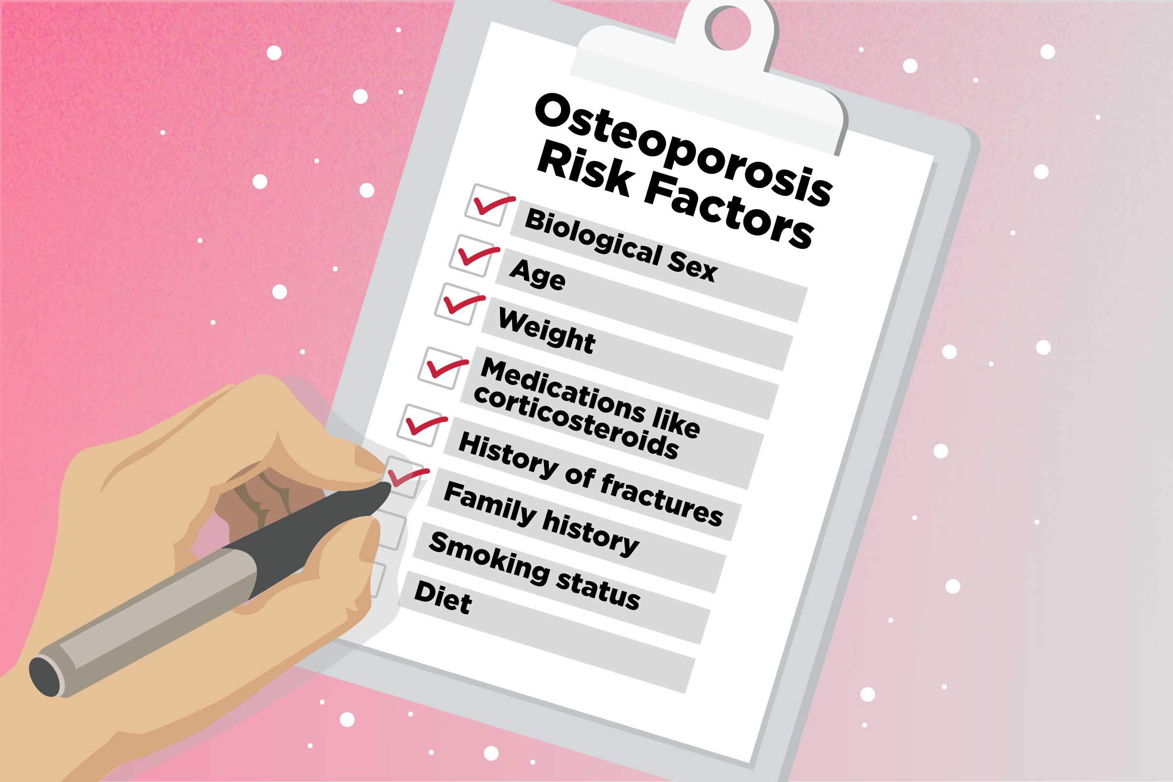
How common are fractures, and who carries the burden?
The burden touches millions each year. Around the world, fractures occur every minute. One in three women over 50 will fracture. One in five men will also fracture. Hip fractures dominate disability and cost. The pattern stresses prevention over late repair. Our editor’s research highlights this steady trend. Numbers keep climbing with aging populations.
Who should be screened, and when should we start?
Screening starts before the first fracture. Women 65 and older need DXA screening. Younger postmenopausal women need risk assessment first. If risk is elevated, order DXA promptly. Men with risk factors also deserve attention. Clinical judgment fills gaps in rigid rules. According to our editor’s research, clear reminders boost uptake. Calendar prompts and recall lists work well.
Which risk tools help you choose the next step?
FRAX estimates 10-year fracture probabilities. It works with or without BMD values. Many systems use fixed intervention thresholds. A 10-year hip risk of 3 percent often prompts therapy. A 20 percent major osteoporotic risk also guides treatment. Thresholds vary across regions and populations. Record the exact calculator and country model used. Consistency improves shared understanding and follow-up.
What labs and imaging feel practical in clinic flow?
Begin with targeted secondary cause screening. Check calcium, vitamin D, and renal function. Consider thyroid and parathyroid tests when indicated. Order DXA for spine and hip as standard. Use VFA when height loss or back pain appears. Repeat DXA on a realistic schedule. Anchor intervals to therapy changes and risk. Keep the testing plan simple and transparent.
How do you talk about lifestyle changes without losing momentum?
Start with everyday habits, not lectures. Tie goals to activities patients love. Walking, dancing, and short resistance sets build bone. Balance training reduces falls and fear. Smoking cessation and alcohol limits help risk. Protein intake supports muscle and bone. Editörümüzün incelemeleri sonucu, brief scripts work best. Simple words outperform dense lists every time.
What about calcium, vitamin D, and protein targets?
Most adults need about 1,000 to 1,200 mg calcium daily. Older adults sit at the upper end. Vitamin D needs remain steady with age. Food should carry most of the load. Supplements fill gaps when diets fall short. Split calcium doses to ease absorption. Pair vitamin D with meals containing fat. Recheck levels when the plan changes.
Which medications matter most, and who should start them?
High-risk patients benefit from antiresorptives first. Bisphosphonates lead many treatment plans. They lower vertebral, nonvertebral, and hip fractures. Denosumab helps when other agents fail or do not fit. Anabolic agents suit very high-risk profiles. Choice depends on risk, comorbidities, and access. As a result of our editor’s reviews, simple decision trees help. Keep the algorithm visible in your workspace.
How long should therapy continue, and what about “holidays”?
Reassess bisphosphonate users after three to five years. Continue if risk remains high. Consider a therapy holiday at lower risk. Monitor BMD and clinical events during any break. Do not interrupt denosumab without a plan. Transition to a bisphosphonate prevents rebound risk. Schedule that transition before the next due dose. Document the dates and the chosen agent.
Why is denosumab discontinuation a special case?
Stopping denosumab can trigger a rebound effect. Bone turnover rises quickly after the last dose. Bone density gains may vanish within months. Vertebral fracture clusters can appear in that window. A bisphosphonate “relay” reduces this rebound. Longer denosumab courses complicate the relay success. Our editor’s research urges early planning here. Do not leave patients between therapies unprotected.
How should you approach glucocorticoid-induced bone loss?
Even low steroid doses raise fracture risk. Risk rises with dose and duration. Start prevention when treatment begins. Add calcium, vitamin D, and exercise advice early. Consider antiresorptives for sustained courses. Reassess if doses change or taper. Build DXA into long-term plans. Treat vertebral fractures aggressively and promptly.
What about men, younger patients, and secondary causes?
Men break bones too, often silently. Secondary causes hide behind vague symptoms. Hypogonadism and alcohol use often play roles. Thyroid and parathyroid disease influence bone turnover. Celiac disease may reduce calcium absorption. Renal disease alters mineral balance. Address root causes alongside medications. Teach patients how fixes reinforce each other.
How do comorbidities shape your medication choice?
Esophagitis complicates oral bisphosphonates. Renal issues limit several agents. Poor adherence weakens weekly schedules. Needle aversion reduces injection acceptance. Upcoming dental surgery affects timing decisions. Prior cardiovascular events influence choices. Very high risk may require anabolic starts. Map the tradeoffs with patients in plain words.
What does a practical monitoring strategy look like?
Tie monitoring to specific questions. Is density stable, rising, or falling? Are there any interval fractures or falls? Are markers telling a consistent story? Is adherence steady by refill counts? Does the patient feel stronger or safer? Adjust plans around these answers. Keep the conversation short and regular.
How should inpatient teams act after a fracture?
Every fracture flags future risk. Post-fracture care must include prevention steps. Order DXA and risk estimates before discharge. Start therapy where criteria are clear. Book follow-up before the patient leaves. Communicate with primary care in writing. Fracture liaison pathways raise adherence. This closes the loop and saves lives.
Where do anabolics and treatment sequencing fit today?
Very high-risk profiles benefit from anabolics first. Romosozumab can be followed by antiresorptives. Sequencing preserves gains and reduces incidents. Use sequences where fracture risk is imminent. Insurance and access shape real choices. Set expectations before starting the path. According to our editor’s research, clarity helps adherence. Patients stay when they see the map.
How do you turn lifestyle advice into repeatable routines?
Write goals in the patient’s own words. Attach actions to daily anchors like meals. Encourage short resistance sets at home. Layer balance work into walking plans. Celebrate small wins at each visit. Limit caffeine and excess salt gradually. Keep alcohol within safer bounds. Bring family into fall-proofing the home.
What nutrition messages actually stick in real life?
Food first remains your north star. Calcium-rich choices should headline meals. Vitamin D needs steady sources year-round. Protein supports muscle and bone repair. Hydration helps training and daily energy. Avoid extreme restrictions that undercut intake. Plan snacks for clinic days and travel. Simple swaps outperform strict diets over time.
What does a lean clinic workflow look like?
Create a standing osteoporosis slot each week. Batch FRAX entries before the visit. Preload labs and prior DXAs into notes. Use smart phrases for counseling scripts. Align refill dates with follow-up windows. Track denosumab renewal dates precisely. Assign a navigator for fracture calls. Review two charts daily for quality checks.
Editor’s quick notes that keep teams aligned
According to our editor’s research, checklists beat memory. Use a one-page algorithm at triage. Color-code thresholds for ease of use. Document the exact FRAX model each time. Record the holiday end date when you start it. Book the bisphosphonate relay before denosumab ends. Print a home exercise card for every patient. Review progress in three sentences or fewer.
A simple conversation map for patients in six steps
Begin with why bones matter now. Share their personal risk in numbers. Offer two clear lifestyle actions today. Explain one medication option with benefits. Discuss likely side effects and monitoring. Agree on the next check-in date. End with a written plan for the fridge. Small, steady steps keep people moving.
Key takeaways for busy internists
Screen on time and act on numbers. Pair FRAX with clinical judgment. Start antiresorptives for high risk. Plan therapy duration from day one. Never stop denosumab without a relay. Train balance and strength every week. Fuel bones with calcium, vitamin D, and protein. Close the loop after every fracture.